E coli gram stain arrangement 267109-Can e coli be gram positive
Table for Staining method Stain Used to Procedure Results Bacteria Negative Identify cell morphology and size 1 Nigrosin, Acid fuchsin, Congo red Dark background, light cells S aureus Simple Identify cell morphology and arrangement 1 Methylene blue, Crystal violet All cells one color B cereus E coli Differential Gram DifferentiateGram Staining of Micrococcus luteus,Escherichia coli, and Serratia marcescens INTRODUCTION In 14, a Danish botanist named Hans Christian Gram developed the popular staining technique known as the "gram stain"His background in studying plants at the University of Copenhagen introduced him to observing different tissues under a microscope, and thus propelled his interest in pharmacologyGramstain Negative = 0, Positive = 1, Indeterminate = 2 Found in human microbiome Microbes that live anywhere in the human body and are not pathogenic to humans (ie capable of causing human disease) No = 0, Yes = 1 Plant pathogen Does the species causes disease in plants?

Virtual Lab 2 tc
Can e coli be gram positive
Can e coli be gram positive-C Gram staining can be used to differentiate intestinal normal microbiota from pathogens—for example differentiating E coli from Salmonella enterica D In most cases, Gram staining is not sufficient to identify an organism—for example, Grampositive staphylococci on skin may be either S aureus or S epidermidisEscherichia coli, often abbreviated E coli, are rodshaped bacteria that tend to occur individually and in large clumps E coli bacteria have a single cell arrangement, according to Schenectady County Community College E coli is a gramnegativ


Lab 1
Note the single bacilli which are characteristic of E coli 1000× Staining microbial cells is important because without stain, most bacterial cells are extremely difficult to see Staining allows them to be seen so that observations as to their morphology (ie, individual cell shape) and arrangement (ie, how the cellsSee more pathogens and parasites on our channel & please subscribe!Note the single bacilli which are characteristic of E coli 1000× Staining microbial cells is important because without stain, most bacterial cells are extremely difficult to see Staining allows them to be seen so that observations as to their morphology (ie, individual cell shape) and arrangement (ie, how the cells
Free YouTube Audio Library Music was used Carol of the Bells by Quincas MoreiraDATA SET Research the following bacterial species and determine the expected results for gram staining, arrangement, capsule staining, and spore staining Gram Stain Arrangement Spore Stain Capsule Stain Escherichia coli K1 Bacillus subtilis Staphylococcus epidermidis Clostridium difficile Mycobacterium leprae Micrococcus luteus Neisseria meningitidesEscherichia coli (/ ˌ ɛ ʃ ə ˈ r ɪ k i ə ˈ k oʊ l aɪ /), also known as E coli (/ ˌ iː ˈ k oʊ l aɪ /), is a Gramnegative, facultative anaerobic, rodshaped, coliform bacterium of the genus Escherichia that is commonly found in the lower intestine of warmblooded organisms (endotherms) Most E coli strains are harmless, but some serotypes (EPEC, ETEC etc) can cause serious food
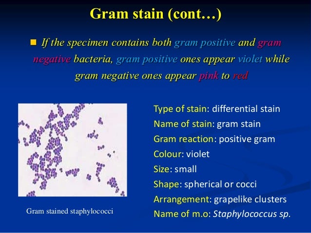
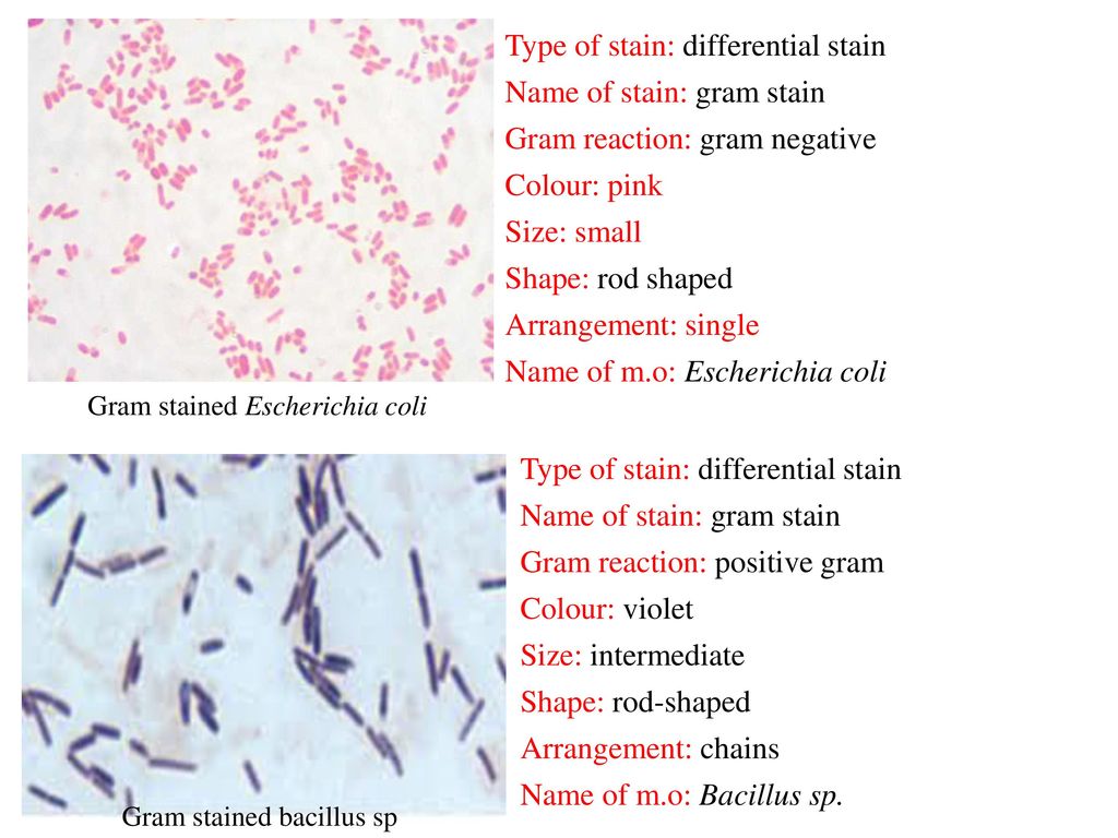
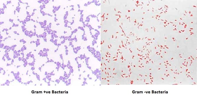
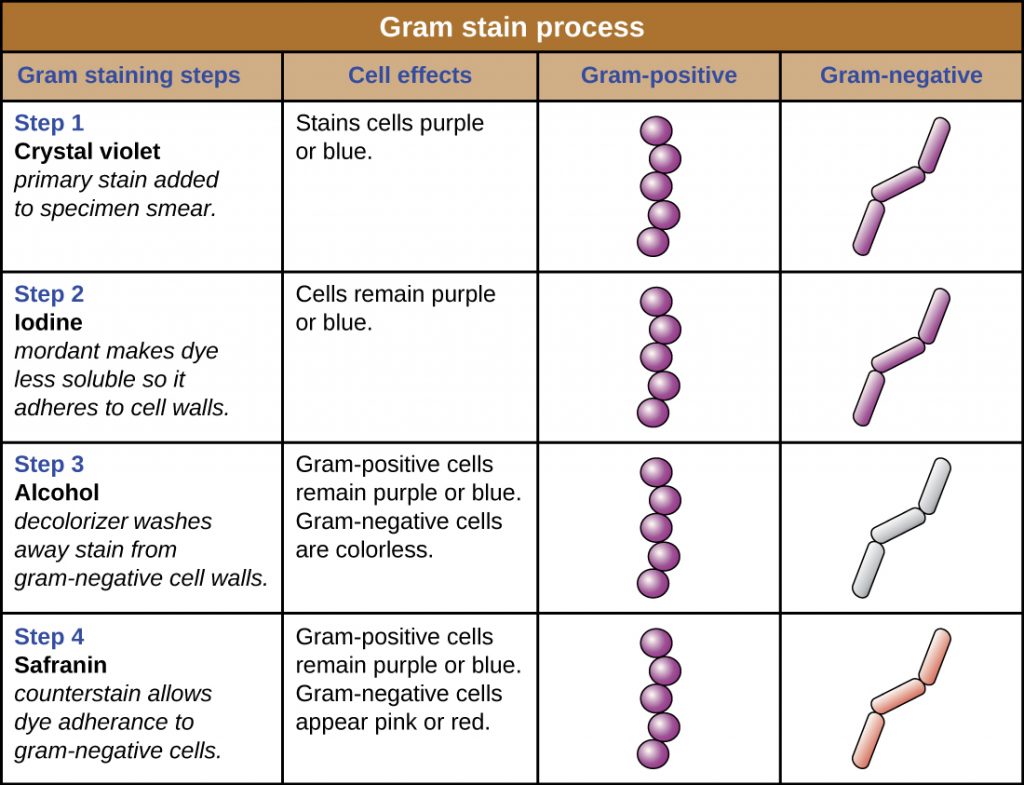
Gram positive bacteria Stain dark purple due to retaining the primary dye called Crystal Violet in the cell wall Example Staphylococcus aureus Fig Gram positive bacteria 2 Gram negative bacteria Stain red or pink due to retaining the counter staining dye called Safranin Example Escherichia coli Fig Gram negative bacteriaHowever, in Gramnegative bacteria, like E coli, the outer membrane blocks vancomycin, which has precluded the use of vancomycin to study these cells We were able to study the effects of vancomycin on E coli by using an imp4213 strain that contains a mutation in the outermembrane protein Imp that perturbs LPS biosynthesisE coli stains Gramnegative because its cell wall is composed of a thin peptidoglycan layer and an outer membrane During the staining process, E coli picks up the color of the counterstain safranin and stains pink


Solved 2 10mm Figure 1 Gram Stain Bacteria Are Escherich Chegg Com


Q Tbn And9gcqkye60ou Johpr02n Mbv1fferrjpdh Lnct7ymdf5qhyia1ld Usqp Cau
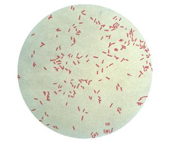
E coli in gram stain E coli in Gram stain showing gram negative rods having size of about µm long and diameter as shown above picture Introduction of Escherichia coli E coli is a Gram negative, aerobe and facultative anaerobic, rodshaped bacteria Optimal temperature for growth is 3617°C with most strains growing over the range 1844 °CEscherichia coli Four different strains of Escherichia coli on Endo agar with biochemical slope Glucose fermentation with gas production, urea and H 2 S negative, lactose positive (with exception of strain D "late lactose fermenter";List at least 3 differences between gram positive and gram negative bacteria Would it be useful to perform a gram stain on a mixed culture?


Solved Simple Stain Q 4 Make Drawings Of Your Simple St Chegg Com


Bacterial Staining
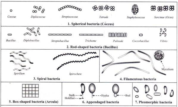
Shape – Escherichia coli is a straight, rod shape (bacillus) bacterium Size – The size of Escherichia coli is about 1–3 µm × 04–07 µm (micrometer) Arrangement Of Cells – Escherichia coli is arranged singly or in pairs Motility – Escherichia coli is a motile bacterium Some strains of E coli are nonmotileNo Can Produce Endospores?DATA SET Research the following bacterial species and determine the expected results for gram staining, arrangement, capsule staining, and spore staining Gram Stain Arrangement Spore Stain Capsule Stain Escherichia coli K1 Bacillus subtilis Staphylococcus epidermidis Clostridium difficile Mycobacterium leprae Micrococcus luteus Neisseria meningitides


Lab 1


Gram Stain Wikipedia
Gram Staining Now that the slide has been heat fixed, you may now begin to stain the organism This is started by applying a few drops of crystal violet for 30 seconds, then rinsing with water for 5 seconds Next, cover the slide with Gram's Iodine and let sit for a full minute before rinsing with water for another 5 secondsBio 112 Abstract Escherichia coli, Bacillus subtilis and Staphylococcus epidermidis were analyzed for this lab activity to determine their Gram Stain After the multilayered Gram Stain procedure each bacteria were classified as Grampositive or Gramnegative depending on their cell walls staining color The results showed that E coli stained pink and classified as GramnegativeThe purpose of this experiment was to determine the Gram stain of the certain slant cultures we used in class, Escherichia coli and Staphylococcus aureus, under a microscope Since it is the most important staining procedure in microbiology, it is used to distinguish between grampositive and gramnegative organisms


Laboratory Perspective Of Gram Staining And Its Significance In Investigations Of Infectious Diseases Thairu Y Nasir Ia Usman Y Sub Saharan Afr J Med


Gram Staining Principle Procedure And Results Msc Sarah Ahmed Ppt Download
In this paper, patches of an E coli cell wall were made using a circumferential layered model, supported by ECT imaging of both Gramnegative and Grampositive sacculi , , plausibility arguments based on the thickness and glycan strand length , and the average peptidepeptide angle measured above (see Fig 2B) The patches were constructedEscherichia coli Escherichia coli (Ecoli) is a common gramnegative bacterial species that is often one of the first ones to be observed by students Most strains of Ecoli are harmless to humans, but some are pathogens and are responsible for gastrointestinal infections They are a bacillus shaped bacteria that has a very fast growth (theyDescribe the Gram reaction, cell shape and arrangement (if there is one) for the following known organisms viewed in the lab E coli (15 marks) Ecoli is a gramnegative bacteria and its shape is rods and arrangement is scattered and are singles/pairs S epidermidis (15 marks) S epidermidis is a grampositive bacteria and its shape and


E Coli Gram Stain Introduction Principle Procedure And Result Interpret


Gram Staining Principle Procedure Interpretation Examples And Animation
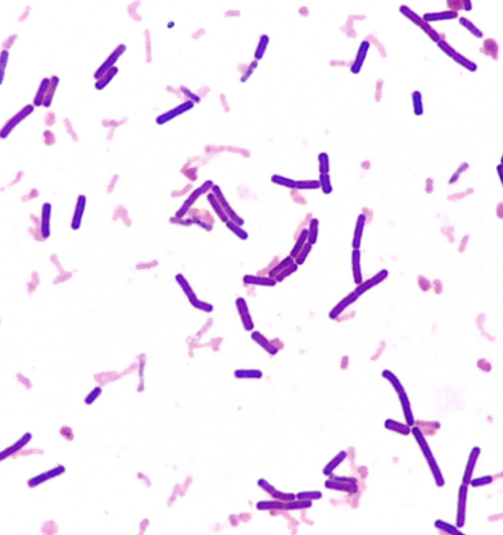
On staining, E coli appear as nonsporeforming, Gramnegative rodshaped bacterium Routine urine cultures should be plated using calibrated loops for the semiquantitative method Note The most commonly used criterion for defining significant bacteriuria is the presence of ⩾10 5 CFU per milliliter of urineWe have cultures of E coli and Bacillus for you to gram stain This will give you gram and gram – controls to check your procedure against You can use 2 slides, 1 for each bacterium, or you can divide one slide in half and smear each bacterium on the divided slide There are also prepared, gram stained slides of bacteria of different shapes and sizes of bacteria to look atDiffusely adherent E coli (DAEC) Gram Stain E coli is described as a Gramnegative bacterium This is because they stain negative using the Gram stain The Gram stain is a differential technique that is commonly used for the purposes of classifying bacteria The staining technique distinguishes between two main types of bacteria (gram


E Coli Gram Stain Page 6 Line 17qq Com


Biol 315l Practical Flashcards Quizlet
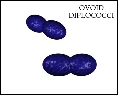
So far as the arrangement is concerned, it may Paired (diplo), Grapelike clusters (staphylo) or Chains (strepto) In shape they may principally be Rods (bacilli), Spheres (cocci), and Spirals (spirillum)On Endo agar it looks like lactose negative)All four strains are mannitol positive (best seen in fig D), cellobiose negative (strains A, B)Escherichia coli, often abbreviated E coli, are rodshaped bacteria that tend to occur individually and in large clumps E coli bacteria have a single cell arrangement, according to Schenectady County Community College E coli is a gramnegative bacillus bacteria that can live with or without oxygen Escherichia coli is normally found amongst the gut flora of the human intestinal tract and is nonpathogenic


Solved Gram Stain Results Below Are Images Of Bacteria I Chegg Com


Morphology Of Bacterial Cells
E coli are Gramnegative bacteria, meaning that they do not retain the crystal violet stain commonly used to differentiate bacteria Their status as Gramnegative bacteria is due to their thin cell walls E coli has cell walls made out of two thing peptidoglycan layers, an inner and outer membraneThe staining technique distinguishes between two main types of bacteria (gram positive and gram negative) by imparting color on the cells Being Gramnegative bacteria, E coli have an additional outer membrane that is composed of phospholipids and lipopolysaccharides2 Morphology and Staining of Escherichia Coli E coli is Gramnegative straight rod, 13 µ x 0407 µ, arranged singly or in pairs (Fig 281) It is motile by peritrichous flagellae, though some strains are nonmotile Spores are not formed Capsules and fimbriae are found in some strains 3 Cultural Characteristics of Escherichia Coli


Solved After The Correct Gram Staining Process A Escheri Chegg Com


Module 7 8 10 Gram Stain Acid Fast Endospore Growth Characteristics Flashcards Quizlet
It is estimated that % of the human population are longterm carriers of S aureusWhat color is E coli when gram stained?2 Morphology and Staining of Escherichia Coli E coli is Gramnegative straight rod, 13 µ x 0407 µ, arranged singly or in pairs (Fig 281) It is motile by peritrichous flagellae, though some strains are nonmotile Spores are not formed Capsules and fimbriae are found in some strains 3 Cultural Characteristics of Escherichia Coli


140mic General Microbiology Ppt Download


Experiment 4a Lab04 Virtual Edge Molb 21 College Of Agriculture And Natural Sciences
What are the gram reaction, shape, and arrangement of E coli?Gramstain Grampositive cocci and Gramnegative bacilli Microscopic appearance Cocci in grapelike clusters (Saureus) and bacilli(Ecoli) Clinical significance of Saureus Frequently found as part of the normal skin flora on the skin and nasal passages;Arrangement streptobacilli Escherichia coli (E coli) Gram negative Gram negative (pink from gram stain);


Pin On Staphylococcus Epidermidis Gram Stain


Bacterial Staining
Diplobacilli (two rods connected in middle) and bacillus (singular rods) 2 diseases caused by Escherichia coli non pathogenic therefore not disease causingEscherichia coli Gram Stain Reaction Gram negative (pink) Bacterial Cell Shape rod Bacterial Cell Arrangement singles Acidfast?The gram stain was devised by histologist Christian Gram as a method of Bacteria sometime show characteristic cellular arrangement or grouping Peritrichous Escherichia coli


Q Tbn And9gcsopf3keyx4bd1kebrm Fh25hgyq4wrk8z 163fpfmbxfw Uw Usqp Cau


Morphology Of Bacterial Cells
Escherichia coli (E coli) as a Model Organism or Host Cell 4 Growth Requirements of E coli and Auxotrophs 3 Actinobacteria Definition & Characteristics 415Gram stain results determine if the organism is gramnegative, but findings do not distinguish among the other aerobic gramnegative bacilli that cause similar infectious diseases E coli is aArrangement streptobacilli Escherichia coli (E coli) Gram negative Gram negative (pink from gram stain);


Gram Stain Arrangement Page 1 Line 17qq Com
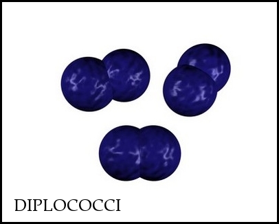


Morphology Of Bacterial Cells
Diplobacilli (two rods connected in middle) and bacillus (singular rods) 2 diseases caused by Escherichia coli non pathogenic therefore not disease causing1 E coli stained with crystal violet @ 100xTM It is not possible to see individual bacteria at this magnification, but viewing at 100XTM is a necessary step in focusing to scope to see at higher magnifications 2 E coli stained with crystal violet @ 1000xTM1 Escherichia coli a Fix a smear of Escherichia coli to the slide as follows 1First place a small piece of tape at one end of the slide and label it with the name of the bacterium you will be placing on that slide 2Using the dropper bottle of deionized water found in your staining rack, place 1/2 of a normal sized drop of water on a clean slide by touching the dropper to the slide
/gram-positive-staphylococcus-aureus-bacteria-541802136-57979cca5f9b58461f26eccc.jpg)


Gram Stain Procedure In Microbiology


Genotypic And Phenotypic Studies Of Murine Intestinal Lactobacilli Species Differences In Mice With And Without Colitis Applied And Environmental Microbiology
For instance, whereas E coli bacteria range between 11 and 15 um in diameters, B anthracis range between 10 and 12um while B subtilis range between 025 and 10um in diameter They also vary in length when compared to each otherE coli S epidermidis Note Escherichia coli is a tiny pink (Gram) rod Staphylococcus epidermidis is a purple (Gram) sphere or coccus Draw a picture of a typical microscopic field and identify both Escherichia coli and Staphylococcus epidermidis Record this in the results section for this lab Colored pencils are available throughout the room1 E coli stained with crystal violet @ 100xTM It is not possible to see individual bacteria at this magnification, but viewing at 100XTM is a necessary step in focusing to scope to see at higher magnifications 2 E coli stained with crystal violet @ 1000xTM


Morphology Culture Characteristics Of Escherichia Coli E Coli


416pht Lab 2 Gram S Stain Acid Fast Stain Spore Stain Ppt Video Online Download
Name the dye that gives it this color To what cell structure do the 2 dyes bind?1 E coli stained with crystal violet @ 100xTM It is not possible to see individual bacteria at this magnification, but viewing at 100XTM is a necessary step in focusing to scope to see at higher magnifications 2 E coli stained with crystal violet @ 1000xTMNo = 0, Yes = 1 Animal pathogen Does the species causes disease


Colony Characteristics Of E Coli Sciencing
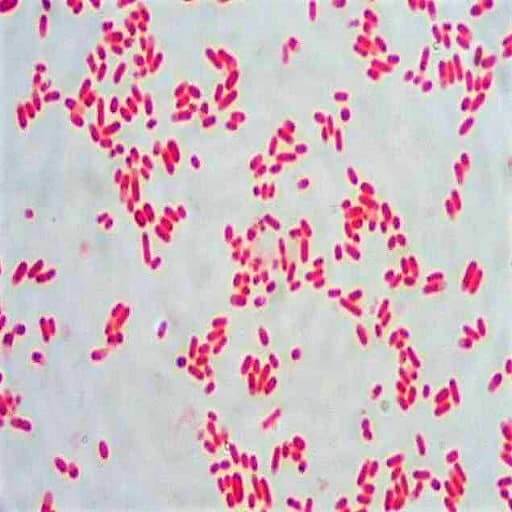


Morphology Culture Characteristics Of Escherichia Coli E Coli
E coli are classified as facultative anaerobes, which means that they grow best when oxygen is present but are able to switch to nonoxygendependent chemical processes in the absence of oxygen E coli bacteria are gramnegative, so they stain pink in a gram test E coli bacteria were first identified by bacteriologist Dr Escherich in 15The Gram stain is the most widely used staining procedure in bacteriology Trypticase Soy agar plate cultures of Escherichia coli (a small, Gramnegative bacillus) is Grampositive or Gramnegative when microscopically viewing a Gram stain preparation and state the shape and arrangement of the organismGram Staining Now that the slide has been heat fixed, you may now begin to stain the organism This is started by applying a few drops of crystal violet for 30 seconds, then rinsing with water for 5 seconds Next, cover the slide with Gram's Iodine and let sit for a full minute before rinsing with water for another 5 seconds


Q Tbn And9gcrnromkd6ojdipvjbbunntiiociojvtit2bpoe0v2w9ohdxp0lr Usqp Cau


Solved Lab Module 6 Gram Stain Lab Report Name Using T Chegg Com
Staphylococci (staphylococcus aureus)gram positive, spherical bacteris that have developed antibiotic resistant strains, 400x gram stain stock pictures, royaltyfree photos & images microscopic view of bacterial pneumonia bacterial pneumonia is a type of pneumonia caused by bacterial infection gram stain stock illustrationsGram Staining of Micrococcus luteus,Escherichia coli, and Serratia marcescens INTRODUCTION In 14, a Danish botanist named Hans Christian Gram developed the popular staining technique known as the "gram stain"His background in studying plants at the University of Copenhagen introduced him to observing different tissues under a microscope, and thus propelled his interest in pharmacology


Escherichia Coli Morphology Mixed Rods Gram Stain Gram Negative Bacilli Growth Characteristics Dubai Khalifa


Solved 1 Identify The Morphology Morphological Arrangem Chegg Com


What Is The Cell Morphology Of Escherichia Coli Quora


Gram Positive Bacteria Microbiology
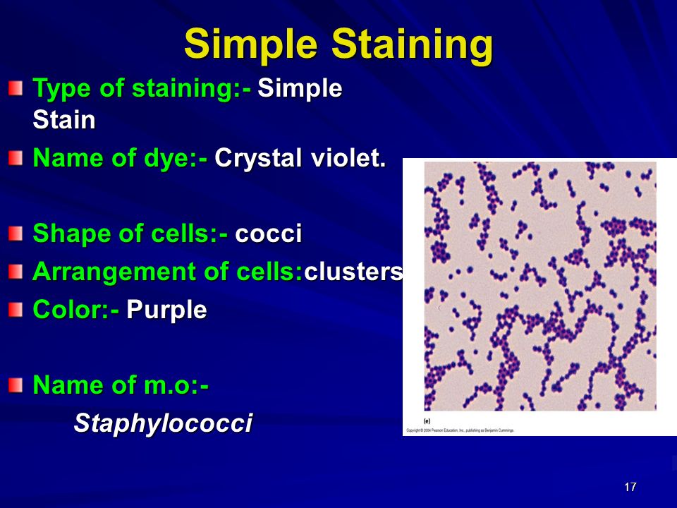


Staining Ppt Video Online Download


Firmicutes Wikipedia


Jn Tm6z15kb1m


Microbiology Lab Test 2 Flashcards Quizlet


E Coli Gram Stain Mikrobiologiya
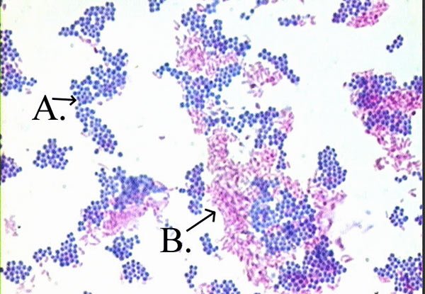


Acid Fast Stain Principle Procedure Interpretation And Examples


Solved B Subtilis Stain Crystal Violet E Coli Stain Chegg Com
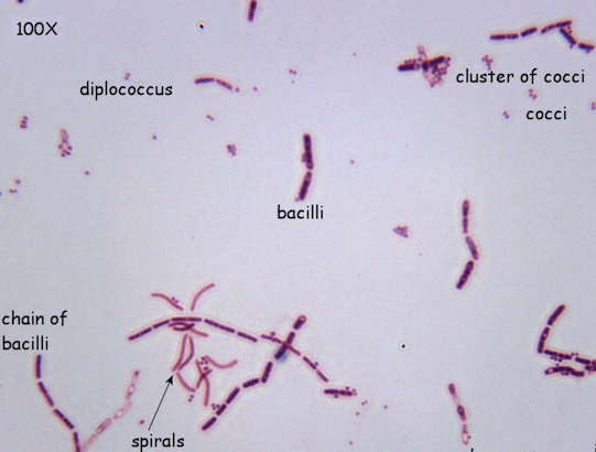


16 Simple Stain Biology Libretexts
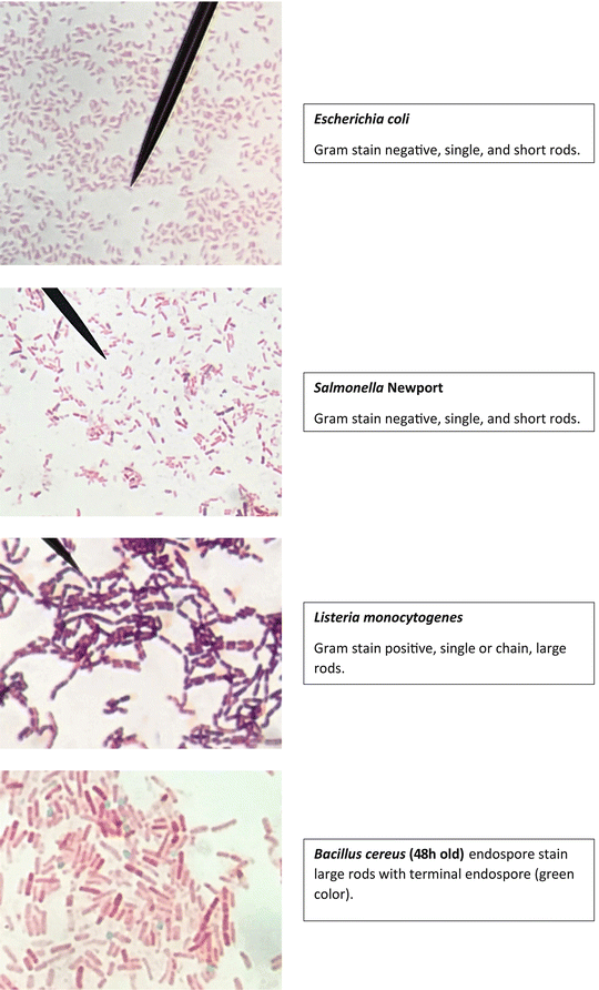


Staining Technology And Bright Field Microscope Use Springerlink
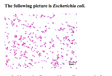


Solved Lab Module Staining Study Help Instructions Rea Chegg Com


Bacteria Exercise Flashcards Quizlet


Lab 3 The Nature Of Bacterial Cell Walls Studocu


Biol 230 Lecture Guide Gram Stain Of A Mixture Of Gram Positive And Gram Negative Bacteria
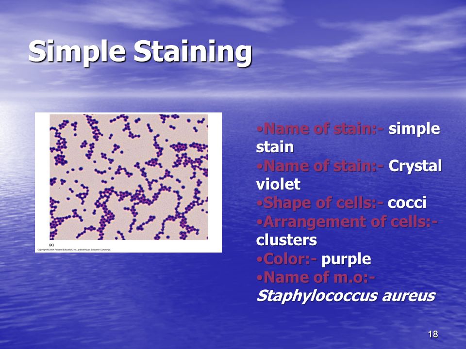


226pht Lab 2 Gram Staining Ppt Video Online Download


Solved 1 Identify The Morphology Morphological Arrangem Chegg Com


Gram Stain By Myriam Feldman Issuu


Virtual Lab 2 tc


Nm6wwwurhlx8ym


Different Size Shape And Arrangement Of Bacterial Cells


What Is The Escherichia Coli S Shape And Arrangement Quora


Colony Characteristics Of E Coli Sciencing


Www Mccc Edu Hilkerd Documents Bio1lab3 Exp 4 Pdf


Pseudomonas Aeruginosa Microbewiki


Www Mccc Edu Hilkerd Documents Bio1lab3 Exp 4 000 Pdf


Everything Micro


Print Micro Exam 2 Flashcards Easy Notecards


Www Mccc Edu Hilkerd Documents Bio1lab3 Exp 4 Pdf


Laboratory Perspective Of Gram Staining And Its Significance In Investigations Of Infectious Diseases Thairu Y Nasir Ia Usman Y Sub Saharan Afr J Med


2 4 Staining Microscopic Specimens Microbiology Canadian Edition


Gram Stain Microbiology Lab


Gram Stain Microbiology Lab
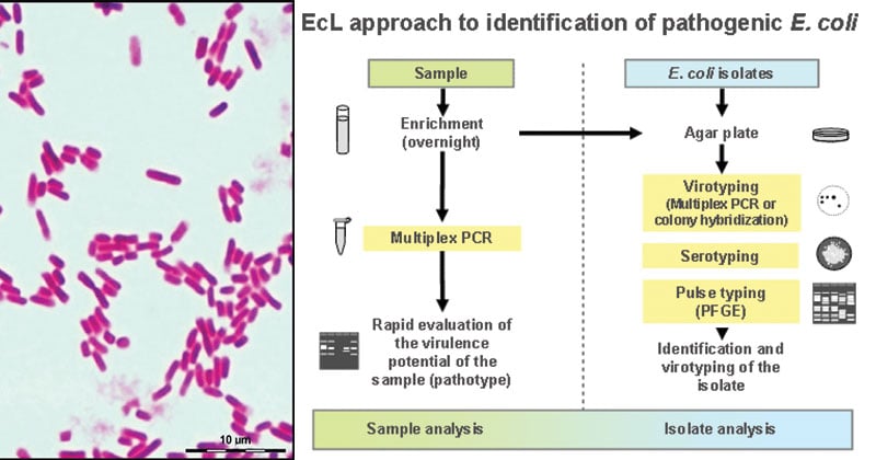


Escherichia Coli E Coli An Overview Microbe Notes


Differential Staining Techniques Microbiology A Laboratory Experience


Gram Stain Staphylococcus Aureus E Coli Combined In Same Organ Control Histology Slides


Microbiology Microbiological Laboratory Systematics Of Microorganisms Morphology Of Microorganims Online Presentation
/gram_positive_vs_negative-5b7f26d2c9e77c005746fbd7.jpg)


Gram Positive Vs Gram Negative Bacteria


72 Microbiology Ideas Microbiology Microbiology Lab Microbiology Study


E Coli Gram Stain Page 1 Line 17qq Com


Www Mccc Edu Hilkerd Documents Bio1lab3 Exp 4 Pdf


Biol 230 Lab Manual Lab 1


E Coli Gram Stain Page 1 Line 17qq Com


Practice Staining


Solved Part I Observe The Gram Stain Results And Colonie Chegg Com


Www Mccc Edu Hilkerd Documents Bio1lab3 Exp 4 Pdf


10 Microbiology Ideas Microbiology Microbiology Lab Medical Laboratory


Solved Lab Module 6 Gram Stain Lab Report Name Using T Chegg Com


The Cell Wall


Escherichia Coli E Coli Meaning Morphology And Characteristics
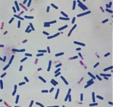


Gram Staining Rules


Module 7 8 10 Gram Stain Acid Fast Endospore Growth Characteristics Flashcards Quizlet


Staphylococcus Aureus And Ecoli Under Microscope Microscopy Of Gram Positive Cocci And Gram Negative Bacilli Morphology And Microscopic Appearance Of Staphylococcus Aureus And E Coli S Aureus Gram Stain And Colony Morphology On Agar Clinical


Bacteria Diversity Of Structure Of Bacteria Britannica


Micrococcus Gram Stain Introduction Principle Procedure And Result Inter


Gram Stain Of E Coli Bacterium A Gram Stain Of Shows Gramnegative Download Scientific Diagram


Identify Shape Arrangement And Gram Stain Flashcards Quizlet


44 Micro Ideas Microbiology Medical Laboratory Microbiology Lab


Solved Unknok 1 What Are The Gram Reaction Morphology Chegg Com


The Gram Stain How To Identify Your Infection Zombie Squad


Bacteria Overview Amboss


E Coli Gram Stain Page 1 Line 17qq Com
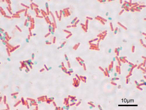


Differential Staining Techniques Microbiology A Laboratory Experience


Www Wiv Isp Be Qml Activities External Quality Rapports Atlas Bacteriology Gram Negative Aerobic And Facultative Rods Pdf
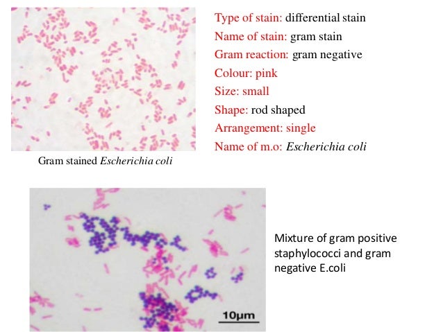


Bacterial Staining
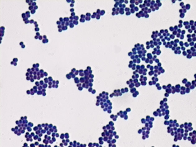


Gram Staining Rules


Gram Stain Of E Coli Bacterium A Gram Stain Of Shows Gramnegative Download Scientific Diagram


Laboratory Perspective Of Gram Staining And Its Significance In Investigations Of Infectious Diseases Thairu Y Nasir Ia Usman Y Sub Saharan Afr J Med


Q Tbn And9gctefycdhnynioi7kgipkpln0ovqbzk1mxi04brg4hvwk5xyhs7g Usqp Cau


What Does An E Coli Bacteria Look Like Under A Microscope Quora


コメント
コメントを投稿